Retinal detachment occurs when the retina separates from the surrounding tissue (the choroid), which supplies it with oxygen and nutrients. When detached, the retina cannot function properly, resulting in vision loss.

The retina is a thin layer of cells lining the inner back wall of the eye. It is the innermost layer of the eye's wall and contains light-sensitive cells called rods and cones, which detect shape, color, and patterns. The retina is supported internally by the jelly-like vitreous, which fills the eyeball behind the lens.
Externally, the retina is attached to the choroid, the middle layer of the eye, which is rich in blood vessels. Nerve fibre from the retina comes together to form the optic nerve, which transmits visual information to the brain.
The most common cause of retinal detachment is age-related shrinkage of the vitreous gel, which can lead to tears in weak areas of the retina. Once a tear or hole forms, fluid can collect beneath it, weakening the adhesion between the retina and the choroid, causing detachment. Eye injuries can also lead to retinal detachment, though this is less common.
Surgery for retinal detachment involves reattaching the retina to the back of the eye and sealing any tears or holes. A retinal specialist will determine the most suitable procedure based on the condition of the eye.
1. Pneumatic Retinopexy
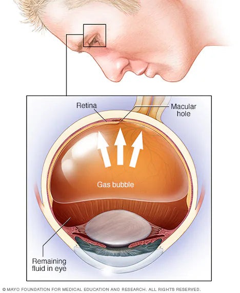
This is the simplest method for repairing detachment, though it may not be suitable for all cases. The surgeon injects a gas bubble into the vitreous cavity and treats the tear(s) with laser or cryotherapy (freezing). The gas bubble presses the retina against the eye wall, while the laser or freezing seals it in place. Post-operative head positioning is crucial for success. The gas bubble dissolves naturally over several days or weeks.
2. Scleral Buckling
Cryotherapy is used to treat the retinal tear, fluid under the retina is drained, and a silicone band is sutured to the sclera (outer eye wall). This band creates an indent that pushes the eye wall back against the retina. The scleral buckle remains permanently unless complications arise.

3. Vitrectomy
During this procedure, the vitreous gel is removed, tears are treated with laser or cryotherapy, and the eye is filled with gas or silicone oil. Correct head positioning post-surgery is essential to allow the retina to reattach. Vision may remain blurry while the gas is absorbed and replaced by natural eye fluid. If silicone oil is used, further surgery is needed to remove it after a few months.
Immediately After Surgery:
During Recovery:
Important Precautions:
If gas was used in the eye, avoid air travel until the gas has fully reabsorbed—this may take up to four weeks.
Seek medical attention immediately if you experience severe pain.
While modern techniques have minimized risks, complications may include:
With appropriate treatment and care, most retinal detachment surgeries are successful in preserving or improving vision.

Frequently asked questions about
What is retinal detachment and how does it affect vision?
Retinal detachment occurs when the retina separates from the choroid, the layer of tissue that supplies it with oxygen and nutrients. When detached, the retina cannot function properly, resulting in partial or complete vision loss in the affected eye. It is a medical emergency that requires prompt surgical treatment to preserve vision.
What causes retinal detachment?
The most common cause of retinal detachment is age-related shrinkage of the vitreous gel inside the eye. As the gel shrinks it can pull on weak areas of the retina and create tears or holes. Once a tear forms, fluid collects beneath the retina and causes it to separate from the eye wall. Eye injuries can also cause retinal detachment though this is less common.
What surgeries are used to repair retinal detachment?
There are three main surgical procedures used to repair retinal detachment. Pneumatic retinopexy involves injecting a gas bubble into the eye to press the retina back against the eye wall. Scleral buckling uses a silicone band sutured to the outside of the eye to push the eye wall against the retina. Vitrectomy removes the vitreous gel and fills the eye with gas or silicone oil. Your retinal surgeon will determine which procedure is most suitable for your specific condition.
Do I need to position my head after retinal detachment surgery?
Yes. Head positioning after retinal detachment surgery is critical for a successful outcome. If a gas bubble was used during surgery — either through pneumatic retinopexy or vitrectomy — you will need to maintain a specific head position to keep the bubble pressing against the area of the retina that needs to heal. Your surgeon will give you exact positioning instructions based on your procedure. Face down positioning is commonly required after vitrectomy for retinal detachment.
How long does recovery take after retinal detachment surgery?
Recovery after retinal detachment surgery varies depending on the procedure used and the severity of the detachment. Discomfort can last several weeks especially if a scleral buckle was used. Blurred vision is common and can take weeks to months — sometimes up to six months — to improve. Vigorous activity should be avoided for several weeks. If a gas bubble was used, air travel must be avoided until the gas has fully reabsorbed which can take up to four weeks.
Can I fly after retinal detachment surgery?
No. If a gas bubble was used during your retinal detachment surgery you must not fly until the gas has completely reabsorbed. Changes in air pressure at altitude cause the gas bubble to expand which can dangerously increase pressure inside the eye. Your surgeon will confirm when it is safe to fly — this typically takes up to four weeks depending on the type of gas used.
What equipment do I need for face down recovery after retinal detachment surgery?
After vitrectomy for retinal detachment you will need face down positioning equipment to maintain the required head position comfortably for the duration of your recovery. The recommended setup includes a face down recovery chair for daytime use, a face down recovery bed for sleeping, a 2-way mirror for watching TV, and a portable seated support for eating and socializing. Having multiple pieces allows you to switch positions throughout the day which reduces discomfort and helps you maintain consistent positioning. RecovRentals provides same-day delivery and setup of medical-grade face down recovery equipment in London, Ontario. Call 519-702-6797 to reserve.
What are the possible complications of retinal detachment surgery?
While modern surgical techniques have significantly minimized risks, possible complications of retinal detachment surgery include cataract formation, raised eye pressure (glaucoma), infection, bleeding into the vitreous cavity, and in rare cases vision loss. Loss of the eye is a very unlikely outcome with modern techniques. With appropriate treatment and care most retinal detachment surgeries are successful in preserving or improving vision. Seek immediate medical attention if you experience severe pain after surgery.
Is retinal detachment surgery successful?
Yes. With appropriate treatment and care most retinal detachment surgeries are successful in preserving or improving vision. Success depends on factors including the severity and location of the detachment, how quickly surgery was performed after detachment occurred, and how consistently the patient follows post-operative instructions including head positioning. Early treatment gives the best chance of a successful outcome.
What is the difference between pneumatic retinopexy, scleral buckling, and vitrectomy for retinal detachment?
Pneumatic retinopexy is the least invasive option — a gas bubble is injected into the eye and laser or freezing seals the tear. It is not suitable for all cases. Scleral buckling involves placing a permanent silicone band around the outside of the eye to push the eye wall back against the retina. Vitrectomy removes the vitreous gel entirely and fills the eye with gas or silicone oil — it is the most common procedure for complex detachments. Your retinal surgeon will recommend the most appropriate procedure based on your specific condition.