
Retinal detachment occurs when the retina separates from the surrounding tissue (the choroid), which supplies it with oxygen and nutrients. When detached, the retina cannot function properly, resulting in vision loss.

The retina is a thin layer of cells lining the inner back wall of the eye. It is the innermost layer of the eye's wall and contains light-sensitive cells called rods and cones, which detect shape, color, and patterns. The retina is supported internally by the jelly-like vitreous, which fills the eyeball behind the lens.
Externally, the retina is attached to the choroid, the middle layer of the eye, which is rich in blood vessels. Nerve fibre from the retina comes together to form the optic nerve, which transmits visual information to the brain.
The most common cause of retinal detachment is age-related shrinkage of the vitreous gel, which can lead to tears in weak areas of the retina. Once a tear or hole forms, fluid can collect beneath it, weakening the adhesion between the retina and the choroid, causing detachment. Eye injuries can also lead to retinal detachment, though this is less common.
Surgery for retinal detachment involves reattaching the retina to the back of the eye and sealing any tears or holes. A retinal specialist will determine the most suitable procedure based on the condition of the eye.
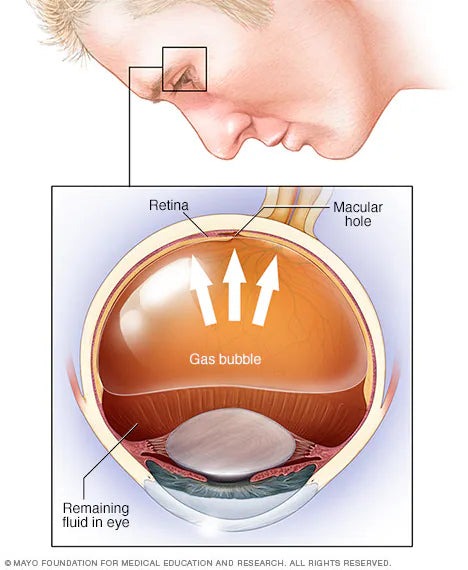
1. Pneumatic Retinopexy
This is the simplest method for repairing detachment, though it may not be suitable for all cases. The surgeon injects a gas bubble into the vitreous cavity and treats the tear(s) with laser or cryotherapy (freezing). The gas bubble presses the retina against the eye wall, while the laser or freezing seals it in place. Post-operative head positioning is crucial for success. The gas bubble dissolves naturally over several days or weeks.
2. Scleral Buckling
Cryotherapy is used to treat the retinal tear, fluid under the retina is drained, and a silicone band is sutured to the sclera (outer eye wall). This band creates an indent that pushes the eye wall back against the retina. The scleral buckle remains permanently unless complications arise.

3. Vitrectomy
During this procedure, the vitreous gel is removed, tears are treated with laser or cryotherapy, and the eye is filled with gas or silicone oil. Correct head positioning post-surgery is essential to allow the retina to reattach. Vision may remain blurry while the gas is absorbed and replaced by natural eye fluid. If silicone oil is used, further surgery is needed to remove it after a few months.
Immediately After Surgery:
During Recovery:
Important Precautions:
If gas was used in the eye, avoid air travel until the gas has fully reabsorbed—this may take up to four weeks.
Seek medical attention immediately if you experience severe pain.
While modern techniques have minimized risks, complications may include:
With appropriate treatment and care, most retinal detachment surgeries are successful in preserving or improving vision.